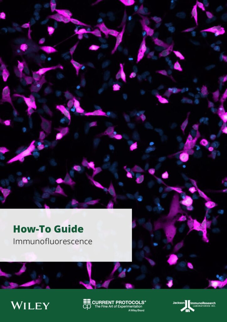
Download this complimentary How-To Guide today!
The guide aims to provide scientists with comprehensive information on immunofluorescence microscopy, enabling you to advance your research in this field.
What you will learn about:
- Microscopy technique selection
- Optimization of imaging parameters
- Troubleshooting fluorescence issues
- Sample preparation and fixation
- Antibody selection and blocking
- Washing and mounting
Download the PDF now.
LOOK INSIDE

More Information
Immunofluorescence microscopy (IF) is a powerful technique used to visualize cellular dynamics by using primary antibodies specific to target proteins in combination with fluorescently labeled secondary antibodies. IF provides insights into the presence, quantity, and subcellular location of target proteins. This technique is applicable to various cell types and requires careful consideration of fixation, permeabilization, and antibody specificity to ensure reliable results.


